Neuronal Wiring Diagram of An Adult Brain
This is my presentation about the Nature paper "Neuronal Wiring Diagram of An Adult Brain". Since I am not a researcher in the field of neuroscience, here is my simple summary of this paper.

Introduction
This paper focuses on studying the brain of Drosopila Melanogaster, which is often refered to as the fruit fly. In the paper, the researchers present a neuronal wiring diagram of a whole brain containing \(5 \times 10^7\) chemical synapses between \(139,255\) neuron reconstructed from an adult female Drosopila melanogaster.

The researchers derive a projectome, which is a map of projections between region, from the connectome and report on tracing of synaptic pathways and the analysis of information flow from inputs to outputs across both hemispheres and between the central brain and optic lobes.
This paper also illustrate how structure can uncover putative circuit mechanisms underlying seasorimotor behaviors.
Data Source
The images of the entire adult female fly brain were previously acquired from the research by Zheng et al.

The authors of this paper previously realigned the electron microscopy images, automatically segmented all neurons in the images, created a computational system that allow interactive proofreading of segmentation, and assembled an online community: Flywire.
- Link to Flywire: https://flywire.ai
- Link to the paper of Flywire: https://www.nature.com/immersive/d42859-024-00053-4/index.html
Motivation and Background
Why we need to map the neuronal wiring of the brain?
Neuroscientists have long recognized that understanding the global connectivity of neurons is the key to understanding how the brain works.
Electron microscopic brain images are applied to chunks of brains to reconstruct local connectivity maps. But nevertheless inadequate for understanding brain function more globally.

And why choose fruit fly as research subject?
Because although small, its brain contain \(10^5\) neurons and \(10^8\) synapses that enable a fly to see, smell, walk and fly. And flies engage in dynamic social interaction, navigate over distance and form long-term memories.
Neurons
Of the 139,255 proofread neurons in FlyWire, 118,501 are intrinsic to the brain, which is defined as the central brain and optic lobes. Intrinsic neurons of the brain make up three-quarters of the adult fly nervous system and amount to 85% of brain neurons.

And brain communicates primarily with itself, and only secondarily with the outside world.
Brain neurons that are not instrinsic can be divided into two categories, afferent and efferent depending on the locations of their cell bodies.
Afferent and Efferent Neurons
-
Afferent (sensory and ascending) neurons: the cell body is outside the brain
-
Efferent (descending, motor and endocrine) neurons: the cell body is contained in the brain
It is generally accurate to think of an afferent neuron as a brain input, and an efferent neuron as a brain output.

Optic Lobes and Central Brain
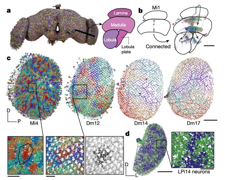
Optic lobe refers to brain structures involved in vision
Visual afferents are by far the most numerous type of sensory input and enter the brain directly rather than through nerves. Of the 118,501 intrinsic neurons, 32,388 are fully contained in the central brain and 77,536 are fully contained in the optic lobes and ocellar ganglia. This number excludes the photorecepter, which are sensory afferent neurons.

The domination of the count by visual areas reflects the nature of Drosophila as a highly visual animal. It shows that vision plays a significant role in neurons and brain.
Synapses and Connections
A Drosophila synapse is generally polyadic, meaning that a single presynapse communicates with multiple target postsynapses. In the research, they define a connection from neuron A to neuron B as the set of synapses from A to B.

Setting a threshold of at least 5 synapses for determining a strong connection is likely to be adequate for avoiding false positive in the dataset while not missing connections.

Projectome

The researchers computed a projectome, which is a neuropil-neuropil matrix, from a synapse-level connectome. Then they weighted neuron projections by the product of the respective number of synapses and normalized the result for every neuron. And they added afferent and efferent neurons to the matrix by calculating the sum of the weighted neuron projections per superclass to and from all neuropils, respectively.


Analysis of Information Flow
Although afferent and efferent neurons make up a small proportion of the brain (13.9% and 1.1%), they connect the brain to the outside world.

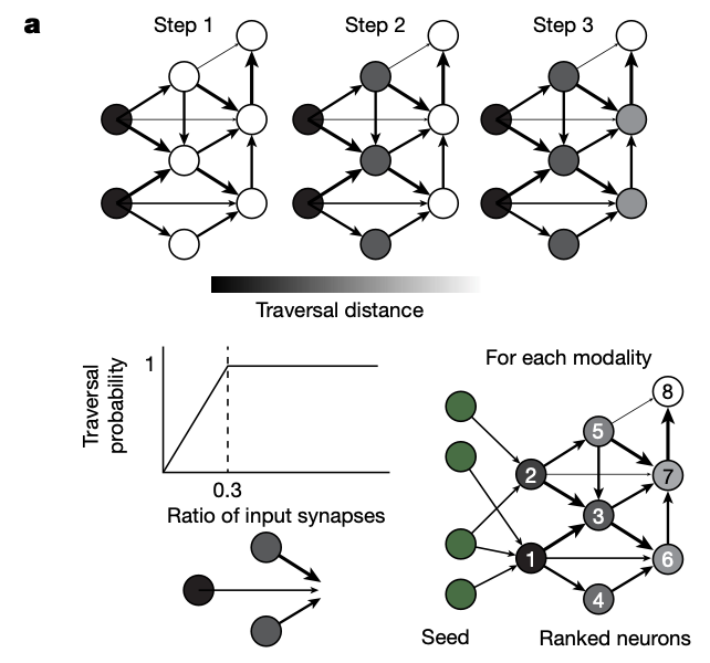
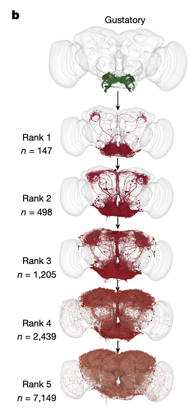
Firstly, the researchers used a probabilistic model to estimate information flow in the connectome, starting from a set of seed neurons. The likelihood of a neuron being traversed increases with the fraction of inputs from already traversed neurons up to an input fraction of 30%, after which traversal is guaranteed.
Next, they measured the flow distance from these starting neurons to all intrinsic and efferent neurons of the central brain.

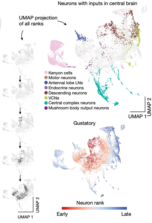
Finally, to visualize information flow for neurons with inputs in the central brain in a common space, they treated the traversal distances starting from each seed population as a neuron embedding and built a uniform manifold approximation and projection (UMAP) from all of these embeddings

Conlustion
Connectome analysis:
- The technologies and open ecosystem reported here set the stage for future large-scale connectome projects in other species
- Has significant benefits for brain research and enables many kinds of studies that were not previously possible using wiring diagrams of portions of the fly brain
Limitation:
- The observed synapse counts underrepresent the actual number of synapses and some connections with few synapses remain undetected
For AI
- The efficient information processing capabilities of the fruit fly brain could provide inspiration for the design of Neural Networks and Large Language Models